
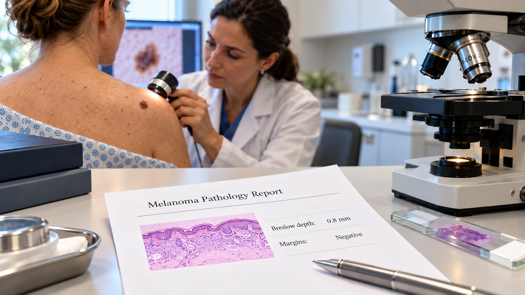
A melanoma pathology report describes findings from a skin biopsy that inform discussions about diagnosis and treatment. Florida’s outdoor lifestyle makes skin health an important conversation in dermatology offices. When a mole changes or a new pigmented lesion raises concern, a dermatologist may recommend biopsy. The pathology report answers a different question from a visual skin examination: what cells and structures are actually present in the sampled tissue? For a confirmed melanoma, the details of that report help the treating team plan the next steps.[1][2]
Key takeaways
- A melanoma pathology report can describe Breslow thickness, ulceration, margins and other relevant findings.
- Pathology information supports staging discussions but does not replace a full clinical evaluation.
- Clinicians should correlate skin biopsy findings with lesion history, examination and any additional assessment.
Melanoma pathology report: why a skin biopsy matters
A dermatologist evaluates a lesion’s appearance, evolution, location and patient history. The widely taught ABCDE warning signs—asymmetry, border irregularity, color variation, diameter and evolution—can help identify lesions that need professional assessment, but they do not establish a diagnosis.[1] Tissue examined microscopically can distinguish a benign melanocytic nevus, an atypical melanocytic proliferation and melanoma. Sometimes inflammatory changes, scar tissue or a different tumor type create a clinically similar appearance.
The biopsy technique also matters. A specimen that includes sufficient lesion depth and representative architecture gives the dermatopathologist a better opportunity to assess the growth pattern and, if melanoma is present, estimate its thickness. The method used for a particular lesion is a clinical decision; not every lesion can or should be approached identically.[2][3]
Melanoma pathology report: findings that deserve attention
Breslow thickness measures the depth of invasion, in millimeters, from a defined epidermal reference point to the deepest melanoma cell. It is not the diameter of the mole and it is not automatically the overall cancer stage. Thickness is an essential element in risk assessment and treatment discussions.[2][3]
Ulceration describes loss of the overlying epidermis attributable to the tumor. Its presence is a relevant staging and prognostic feature. A report may also describe the melanoma subtype, mitotic activity, tumor-infiltrating lymphocytes, microsatellites or other features when applicable.[3]
Peripheral and deep margins indicate whether tumor extends to examined tissue edges. A biopsy with involved margins is not the same as a definitive wide excision; management depends on the diagnosis, sampling and clinical circumstances. In a superficially sampled lesion, an involved deep margin can also limit the precision of the measured thickness.[3]
Why dermatopathology requires clinical correlation
Interpretation is strongest when the pathologist receives the exact lesion site, clinical differential diagnosis, patient history and relevant prior biopsy information. A recurrent pigmented lesion in a scar, for example, may require a different analysis from a new lesion on previously unaffected skin. For unusual melanocytic proliferations, additional tissue levels, immunohistochemistry or a second specialist review may be useful; no single stain resolves every borderline lesion.
For a dermatologist in Orlando, Winter Park or elsewhere in Central Florida, the practical objective is a report that answers the clinical question and clearly communicates uncertainty when it remains. Pathology is one part of care alongside examination, imaging or nodal assessment when indicated.
What happens after the report?
A melanoma diagnosis is followed by an individualized discussion of definitive excision, any need for nodal evaluation and surveillance. Those decisions belong to the treating clinical team and depend on the full report plus patient factors. A pathology report alone cannot determine a person’s treatment plan or prognosis.[2]
Pathology Specialists offers dermatopathology services and case consultation from Orlando. Referring clinicians can contact the local team to discuss specimen submission or a difficult diagnosis; patients should request personalized interpretation from their treating dermatologist.
Information that helps a dermatopathologist interpret the specimen
For referring dermatologists, useful requisition details include the exact body site and laterality, whether a lesion is new or recurrent, the clinical differential diagnosis, biopsy type and relevant previous pathology. A lesion from a nail unit, acral skin or previously treated site may raise a different microscopic differential than one from chronically sun-exposed trunk or facial skin. Securely shared clinical photographs can be valuable when the laboratory’s workflow permits them. The goal is not to influence the diagnosis; it is to prevent loss of context that may determine whether an otherwise ambiguous growth pattern is meaningful. When melanoma is suspected, clinicians should make the collection approach and tissue orientation as informative as clinically feasible.[2][3]
Frequently asked questions
Does an atypical mole mean melanoma?
No. The term atypical may describe changes that are not sufficient for melanoma. The complete diagnosis and clinical context are important.
Is Breslow thickness the same as stage?
No. It is an important measurement used within the broader staging and clinical evaluation process.[3]
Can a skin biopsy be reviewed again?
Yes. When the interpretation is uncertain or treatment depends on a difficult distinction, the referring clinician may request a dermatopathology consultation.
For general educational purposes; individual evaluation and management should be discussed with the treating clinician.